
Advanced Characterization
I have long been fascinated by materials characterization, and in-particular the ability to image atoms and nanoscale phenomena at high spatial resolution. One of the most versatile instruments to achieve this is the scanning transmission electron microscope (STEM).
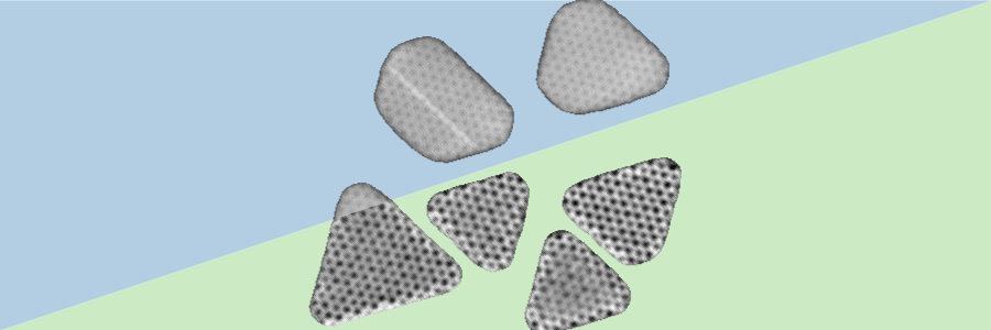
In my PhD work, I have collaborated with experimentalists to directly image moirè superlattices at the interface of 2D/3D materials and investigate phonon localization at the interface between two 2D materials with high-resolution STEM-based techniques.
Currently, as a postdoctoral Miller research fellow, I’m developing computational imaging techniques to image the induced magnetic fields arising in hydrodynamic electron flows with nanoscale resolution, by developing electron ptychography algorithms to jointly-reconstruct the electrostatic and magnetic-vector potentials.